SFB 1192
EM-Facility
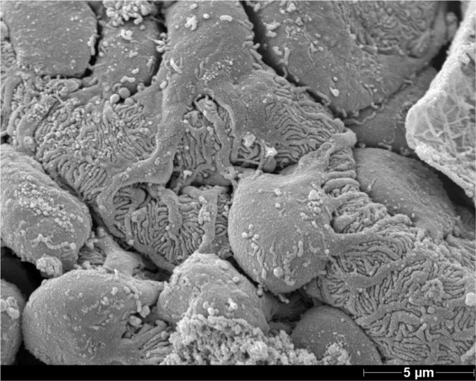
Many of the phenotypes in human kidney disease are only detectable at the ultrastructural level. This holds also true for the model systems of genetic and epigenetic based disturbances of renal development, biology or aging. Our group applies transmission and scanning microscope techniques in human and animal tissue (e.g. mouse, rat, zebrafish, drosophila) in order to detect and characterize subtle ultrastructural alterations e.g. of the glomerular filtration barrier, the actin cytoskeleton or intracellular vesicle formation and tracking at a qualitative and quantitative level. Moreover, pre- and post-embedding immunogold labelling allows us to follow subcellular redistribution of proteins under disease conditions.
We are currently establishing freeze fracture/replica immunogold labelling of kidney tissue and correlative light electron microscopy (CLEM) especially in combination with 3D clarity immunofluorescence imaging in order to broaden our toolbox for the analysis and understanding of renal structural alterations in disease.
Scientifically, the group is currently working on "The role of DNA repair mechanisms on podocyte aging and the consequences of kidney aging on other organs". This study is funded by the DFG and is performed in cooperation with Prof. Tobias Huber (UKE), Prof. Ketan Patel (Reading, UK) und Prof. Jan Hoeijmakers (Rotterdam, NL).

Latest Publications
Procachectic factors link experimental and human chronic kidney disease to skeletal muscle wasting programs.
Solagna F, Tezze C, Lindenmeyer MT, Lu S, Wu G, Liu S, Zhao Y, Mitchell R, Meyer C, Omairi S, Kilic T, Paolini A, Ritvos O, Pasternack A, Matsakas A, Kylies D, Wiesch JSZ, Turner JE, Wanner N, Nair V, Eichinger F, Menon R, Martin IV, Klinkhammer BM, Hoxha E, Cohen CD, Tharaux PL, Boor P, Ostendorf T, Kretzler M, Sandri M, Kretz O, Puelles VG, Patel K, Huber TB.
J Clin Invest. 2021 Jun
ADAM10-Mediated Ectodomain Shedding Is an Essential Driver of Podocyte Damage.
Sachs M, Wetzel S, Reichelt J, Sachs W, Schebsdat L, Zielinski S, Seipold L, Heintz L, Müller SA, Kretz O, Lindenmeyer M, Wiech T, Huber TB, Lüllmann-Rauch R, Lichtenthaler SF, Saftig P, Meyer-Schwesinger C.
J Am Soc Nephrol. 2021 Mar
SRGAP1 Controls Small Rho GTPases To Regulate Podocyte Foot Process Maintenance.
Rogg M, Maier JI, Dotzauer R, Artelt N, Kretz O, Helmstädter M, Abed A, Sammarco A, Sigle A, Sellung D, Dinse P, Reiche K, Yasuda-Yamahara M, Biniossek ML, Walz G, Werner M, Endlich N, Schilling O, Huber TB, Schell C.
J Am Soc Nephrol. 2021 Jan
The proteomic landscape of small urinary extracellular vesicles during kidney transplantation.
Braun F, Rinschen M, Buchner D, Bohl K, Plagmann I, Bachurski D, Richard Späth M, Antczak P, Göbel H, Klein C, Lackmann JW, Kretz O, Puelles VG, Wahba R, Hallek M, Schermer B, Benzing T, Huber TB, Beyer A, Stippel D, Kurschat CE, Müller RU.
J Extracell Vesicles. 2020 Oct
Distinct Modes of Balancing Glomerular Cell Proteostasis in Mucolipidosis Type II and III Prevent Proteinuria.
Sachs W, Sachs M, Krüger E, Zielinski S, Kretz O, Huber TB, Baranowsky A, Westermann LM, Voltolini Velho R, Ludwig NF, Yorgan TA, Di Lorenzo G, Kollmann K, Braulke T, Schwartz IV, Schinke T, Danyukova T, Pohl S, Meyer-Schwesinger C.J
J Am Soc Nephrol. 2020 Jul
Plasminogen deficiency does not prevent sodium retention in a genetic mouse model of experimental nephrotic syndrome.
Xiao M, Bohnert BN, Aypek H, Kretz O, Grahammer F, Aukschun U, Wörn M, Janessa A, Essigke D, Daniel C, Amann K, Huber TB, Plow EF, Birkenfeld AL, Artunc F
Acta Physiol (Oxf). 2020 May
Microbiota-Induced Type I Interferons Instruct a Poised Basal State of Dendritic Cells.
Schaupp L, Muth S, Rogell L, Kofoed-Branzk M, Melchior F, Lienenklaus S, Ganal-Vonarburg SC, Klein M, Guendel F, Hain T, Schütze K, Grundmann U, Schmitt V, Dorsch M, Spanier J, Larsen PK, Schwanz T, Jäckel S, Reinhardt C, Bopp T, Danckwardt S, Mahnke K, Heinz GA, Mashreghi MF, Durek P, Kalinke U, Kretz O, Huber TB, Weiss S, Wilhelm C, Macpherson AJ, Schild H, Diefenbach A, Probst HC.
Cell. 2020 May
The tetraspanin CD9 controls migration and proliferation of parietal epithelial cells and glomerular disease progression.
Lazareth H, Henique C, Lenoir O, Puelles VG, Flamant M, Bollée G, Fligny C, Camus M, Guyonnet L, Millien C, Gaillard F, Chipont A, Robin B, Fabrega S, Dhaun N, Camerer E, Kretz O, Grahammer F, Braun F, Huber TB, Nochy D, Mandet C, Bruneval P, Mesnard L, Thervet E, Karras A, Le Naour F, Rubinstein E, Boucheix C, Alexandrou A, Moeller MJ, Bouzigues C, Tharaux PL.
Nat Commun. 2019 Jul
Novel 3D analysis using optical tissue clearing documents the evolution of murine rapidly progressive glomerulonephritis.
Puelles VG, Fleck D, Ortz L, Papadouri S, Strieder T, Böhner AMC, van der Wolde JW, Vogt M, Saritas T, Kuppe C, Fuss A, Menzel S, Klinkhammer BM, Müller-Newen G, Heymann F, Decker L, Braun F, Kretz O, Huber TB, Susaki EA, Ueda HR, Boor P, Floege J, Kramann R, Kurts C, Bertram JF, Spehr M, Nikolic-Paterson DJ, Moeller MJ.
Kidney Int. 2019 Aug
Compression of morbidity in a progeroid mouse model through the attenuation of myostatin/activin signalling.
Alyodawi K, Vermeij WP, Omairi S, Kretz O, Hopkinson M, Solagna F, Joch B, Brandt RMC, Barnhoorn S, van Vliet N, Ridwan Y, Essers J, Mitchell R, Morash T, Pasternack A, Ritvos O, Matsakas A, Collins-Hooper H, Huber TB, Hoeijmakers JHJ, Patel K.
J Cachexia Sarcopenia Muscle. 2019 Jun
Anaerobic Glycolysis Maintains the Glomerular Filtration Barrier Independent of Mitochondrial Metabolism and Dynamics.
Brinkkoetter PT, Bork T, Salou S, Liang W, Mizi A, Özel C, Koehler S, Hagmann HH, Ising C, Kuczkowski A, Schnyder S, Abed A, Schermer B, Benzing T, Kretz O, Puelles VG, Lagies S, Schlimpert M, Kammerer B, Handschin C, Schell C, Huber TB.
Cell Rep. 2019 Apr
Secretome of adipose-derived mesenchymal stem cells promotes skeletal muscle regeneration through synergistic action of extracellular vesicle cargo and soluble proteins.
Mitchell R, Mellows B, Sheard J, Antonioli M, Kretz O, Chambers D, Zeuner MT, Tomkins JE, Denecke B, Musante L, Joch B, Debacq-Chainiaux F, Holthofer H, Ray S, Huber TB, Dengjel J, De Coppi P, Widera D, Patel K.
Stem Cell Res Ther. 2019 Apr
Project-Team

Project Leader:
PD Dr. Oliver Kretz

